MRI Scans
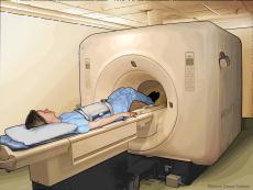
Magnetic resonance imaging (MRI)
uses a large magnet and radio waves to look at organs and structures
inside your body. Health care professionals use MRI scans to diagnose a
variety of conditions, from torn ligaments to tumors. MRIs are very
useful for examining the brain and spinal cord.
During the scan, you lie on a table that slides inside a tunnel-shaped machine. Doing the scan can take a long time, and you must stay still. The scan is painless. The MRI machine makes a lot of noise. The technician may offer you earplugs.
Before you get a scan, tell your doctor if you
During the scan, you lie on a table that slides inside a tunnel-shaped machine. Doing the scan can take a long time, and you must stay still. The scan is painless. The MRI machine makes a lot of noise. The technician may offer you earplugs.
Before you get a scan, tell your doctor if you
- Are pregnant
- Have pieces of metal in your body. You might have metal in your body if you have a shrapnel or bullet injury or if you are a welder
- Have electronic devices in your body, such as a cardiac pacemaker
CT Scans
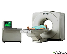
Computed tomography (CT) is a diagnostic procedure that uses special
X-ray equipment to create cross-sectional pictures of your body. CT
images are produced using X-ray technology and powerful computers.
The uses of CT include looking for
The uses of CT include looking for
- Broken bones
- Cancers
- Blood clots
- Signs of heart disease
- Internal bleeding
Intravenous pyelogram
An intravenous pyelogram (IVP) is a special x-ray examination of the kidneys, bladder, and ureters (the tubes that carry urine from the kidneys to the bladder).
How the Test is PerformedAn IVP is done in a hospital radiology department or a health care provider’s office by an x-ray technician.
You will need to empty your bladder immediately before the procedure starts.
The health care provider will inject an iodine-based contrast (dye) into a vein in your arm. A series of x-ray images are taken at different times to see how the kidneys remove the dye and how it collects in your urine.
A compression device (a wide belt containing two balloons that can be inflated) may be used to keep the contrast material in the kidneys.
You will need to remain still during the procedure, which may take up to an hour.
Before the final image is taken, you will be asked to urinate again, to see how well the bladder has emptied.
You can resume your normal diet and medications after the procedure. You should drink plenty of fluids to help remove all the contrast dye from your body.
How to Prepare for the TestAs with all x-ray procedures, tell your health care provider if you:
You must sign a consent form. You will be asked to wear a hospital gown and to remove all jewelry.
How the Test Will FeelYou may feel a burning or flushing sensation in your arm and body as the contrast dye is injected. You may also have a metallic taste in your mouth. This is normal and will quickly disappear.
Some people develop a headache, nausea, or vomiting after the dye is injected.
The belt across the kidneys may feel tight over your belly area.
Why the Test is PerformedAn IVP can be used to evaluate:
Additional conditions under which the test may be performed:
There is low radiation exposure. X-rays are monitored and regulated to provide the minimum amount of radiation exposure needed to produce the image. Most experts feel that the risk is low compared with the benefits.
Pregnant women and children are more sensitive to the risks of radiation.
Considerations Computed tomography (CT) scans have replaced IVP as the main tool for checking the urinary system. CT takes less time to perform and provides additional views of the abdomen, which can help rule out other possible reasons for the patient's symptoms. Magnetic resonance imaging (MRI) is also used to look at the kidneys, ureters, and bladder.
How the Test is PerformedAn IVP is done in a hospital radiology department or a health care provider’s office by an x-ray technician.
You will need to empty your bladder immediately before the procedure starts.
The health care provider will inject an iodine-based contrast (dye) into a vein in your arm. A series of x-ray images are taken at different times to see how the kidneys remove the dye and how it collects in your urine.
A compression device (a wide belt containing two balloons that can be inflated) may be used to keep the contrast material in the kidneys.
You will need to remain still during the procedure, which may take up to an hour.
Before the final image is taken, you will be asked to urinate again, to see how well the bladder has emptied.
You can resume your normal diet and medications after the procedure. You should drink plenty of fluids to help remove all the contrast dye from your body.
How to Prepare for the TestAs with all x-ray procedures, tell your health care provider if you:
- Are allergic to contrast material
- Are pregnant
- Have any drug allergies
You must sign a consent form. You will be asked to wear a hospital gown and to remove all jewelry.
How the Test Will FeelYou may feel a burning or flushing sensation in your arm and body as the contrast dye is injected. You may also have a metallic taste in your mouth. This is normal and will quickly disappear.
Some people develop a headache, nausea, or vomiting after the dye is injected.
The belt across the kidneys may feel tight over your belly area.
Why the Test is PerformedAn IVP can be used to evaluate:
- An abdominal injury
- Bladder and kidney infections
- Blood in the urine
- Flank pain (possibly due to kidney stones)
- Tumors
Additional conditions under which the test may be performed:
- Acute arterial occlusion of the kidney
- Acute bilateral obstructive uropathy
- Acute kidney infection
- Acute unilateral obstructive uropathy
- Bilateral hydronephrosis
- Carcinoma of the renal pelvis or ureter
- Chronic bilateral obstructive uropathy
- Chronic unilateral obstructive uropathy
- Hydronephrosis (swelling of one kidney due to a backup of urine)
- Injury of the kidney and ureter
- Medullary cystic disease
- Polycystic kidney disease
- Reflux nephropathy
- Renal cell carcinoma
- Renal papillary necrosis
- Renovascular hypertension
- Retroperitoneal fibrosis
- Ureterocele
- Wilms tumor
There is low radiation exposure. X-rays are monitored and regulated to provide the minimum amount of radiation exposure needed to produce the image. Most experts feel that the risk is low compared with the benefits.
Pregnant women and children are more sensitive to the risks of radiation.
Considerations Computed tomography (CT) scans have replaced IVP as the main tool for checking the urinary system. CT takes less time to perform and provides additional views of the abdomen, which can help rule out other possible reasons for the patient's symptoms. Magnetic resonance imaging (MRI) is also used to look at the kidneys, ureters, and bladder.
Nuclear Scans
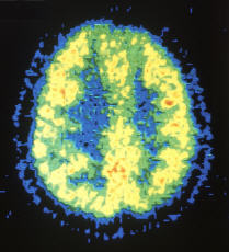
Nuclear scanning uses radioactive
substances to see structures and functions inside your body. Nuclear
scans involve a special camera that detects energy coming from the
radioactive substance, called a tracer. Before the test, you receive the
tracer, often by an injection. Although tracers are radioactive, the
dosage is small. During most nuclear scanning tests, you lie still on a
scanning table while the camera makes images. Most scans take 20 to 45
minutes.
Nuclear scans can help doctors diagnose many conditions, including cancers, injuries and infections. They can also show how organs like your heart and lungs are working.
Nuclear scans can help doctors diagnose many conditions, including cancers, injuries and infections. They can also show how organs like your heart and lungs are working.
PET Scan
A positron emission tomography (PET)
scan is an imaging test that uses a radioactive substance (called a
tracer) to look for disease in the body.
Unlike magnetic resonance imaging (MRI) and computed tomography (CT) scans, which reveal the structure of and blood flow to and from organs, a PET scan shows how organs and tissues are working.
See also:
How the Test is PerformedThe health care provider will inject a small amount of a radioactive material into one of your veins, usually on the inside of the elbow. The substance travels through the blood and collects in organs and tissues.
You'll be asked to wait nearby as the radioactive substance is absorbed by your body. This usually takes about 1 hour.
Then, you will lie down on a table that slides into a tunnel-shaped hole in the center of the PET scanner.
The PET machine detects energy given off by the radioactive substance and a computer changes the results into 3-dimensional pictures. The images are displayed on a monitor for the health care provider to read.
You must lie still during the PET scan so that the machine can produce clear images. How long the test takes depends on what part of the body is being scanned. Today, most PET scans are done with CT scanning.
How to Prepare for the TestYou must sign a consent form before having this test. You will be told not to eat anything for 4 - 6 hours before the PET scan, although you will be able to drink water.
Tell your health care provider if you are pregnant or think you might be pregnant.
Also tell your health care provider about any prescription and over-the-counter medicines that you are taking, because they may interfere with the test.
Be sure to mention if you have any allergies, or if you've had any recent imaging studies using injected dye (contrast).
During the test, you may need to wear a hospital gown. Take off any jewelry, dentures, and other metal objects because they could affect the scan results.
How the Test Will FeelYou will feel a sharp prick when the needle with the radioactive substance is inserted into your vein. You shouldn't feel anything during the actual PET scan.
Why the Test is PerformedA PET scan can reveal the size, shape, position, and some function of organs.
This test can be used to:
For more information see:
Normal ResultsThere are no problems detected in the size, shape, or position of an organ. There are no areas in which the radiotracer has abnormally collected.
What Abnormal Results Mean
RisksThe amount of radiation used in a PET scan is low. It is about the same amount of radiation as in most CT scans. Also, the radiation doesn't last for very long in your body.
However, women who are pregnant or are breastfeeding should let their doctor know before having this test. Infants and babies developing in the womb are more sensitive to the effects of radiation because their organs are still growing.
It is possible, although very unlikely, to have an allergic reaction to the radioactive substance. Some people have pain, redness, or swelling at the injection site.
ConsiderationsIt is possible to have false results on a PET scan. Blood sugar or insulin levels may affect the test results in people with diabetes.
Most PET scans are now performed along with a CT scan. This combination scan is called a PET/CT.
Unlike magnetic resonance imaging (MRI) and computed tomography (CT) scans, which reveal the structure of and blood flow to and from organs, a PET scan shows how organs and tissues are working.
See also:
How the Test is PerformedThe health care provider will inject a small amount of a radioactive material into one of your veins, usually on the inside of the elbow. The substance travels through the blood and collects in organs and tissues.
You'll be asked to wait nearby as the radioactive substance is absorbed by your body. This usually takes about 1 hour.
Then, you will lie down on a table that slides into a tunnel-shaped hole in the center of the PET scanner.
The PET machine detects energy given off by the radioactive substance and a computer changes the results into 3-dimensional pictures. The images are displayed on a monitor for the health care provider to read.
You must lie still during the PET scan so that the machine can produce clear images. How long the test takes depends on what part of the body is being scanned. Today, most PET scans are done with CT scanning.
How to Prepare for the TestYou must sign a consent form before having this test. You will be told not to eat anything for 4 - 6 hours before the PET scan, although you will be able to drink water.
Tell your health care provider if you are pregnant or think you might be pregnant.
Also tell your health care provider about any prescription and over-the-counter medicines that you are taking, because they may interfere with the test.
Be sure to mention if you have any allergies, or if you've had any recent imaging studies using injected dye (contrast).
During the test, you may need to wear a hospital gown. Take off any jewelry, dentures, and other metal objects because they could affect the scan results.
How the Test Will FeelYou will feel a sharp prick when the needle with the radioactive substance is inserted into your vein. You shouldn't feel anything during the actual PET scan.
Why the Test is PerformedA PET scan can reveal the size, shape, position, and some function of organs.
This test can be used to:
- Check brain function
- Diagnose cancer, heart problems, and brain disorders
- See how far cancer has spread
- Show areas in which there is poor blood flow to the heart
For more information see:
Normal ResultsThere are no problems detected in the size, shape, or position of an organ. There are no areas in which the radiotracer has abnormally collected.
What Abnormal Results Mean
- Abnormal heart function
- Abnormal size, shape, or position of an organ
- Alzheimer's disease
- Cancer or tumors
- Change in organ function
- Infection
RisksThe amount of radiation used in a PET scan is low. It is about the same amount of radiation as in most CT scans. Also, the radiation doesn't last for very long in your body.
However, women who are pregnant or are breastfeeding should let their doctor know before having this test. Infants and babies developing in the womb are more sensitive to the effects of radiation because their organs are still growing.
It is possible, although very unlikely, to have an allergic reaction to the radioactive substance. Some people have pain, redness, or swelling at the injection site.
ConsiderationsIt is possible to have false results on a PET scan. Blood sugar or insulin levels may affect the test results in people with diabetes.
Most PET scans are now performed along with a CT scan. This combination scan is called a PET/CT.
DTPA Scan
1. What is a Nuclear Medicine Renal Scan?
The kidneys filter the blood to remove waste substances such as urea (a nitrogen compound) and salt. The body discharges these wastes mixed in water as urine. The fluid is collected in the kidneys and discharged through the ureters which join the kidneys to the bladder. The top of the ureter is called the renal pelvis and this joins the kidney to the ureters.
In a Nuclear Medicine (NM) Renal Scan, images are made of the delivery of fluid into the kidneys via the bloodstream, concentration of wastes in the kidney and excretion or flow from the kidneys through the ureters and filling of the bladder.
A Nuclear Medicine Renal Scan can be performed with 2 different substances - DTPA or MAG3. DTPA is the radiopharmaceutical used in a DTPA Renal Scan, but sometimes the nuclear medicine specialist will decide that another radiopharmaceutical called MAG3 should be used. These radiopharmaceuticals are similar, but MAG3 gives significantly better images in some patients, particularly very young children and those patients with poor kidney function. The descriptions and explanations below for a DTPA renal scan apply also to a MAG3 renal scan. A Nuclear Medicine DTPA or MAG3 Renal Scan is performed to look at the blood supply, function and excretion of urine from the kidneys. The test can find out what percentage each kidney contributes to the total kidney function. A DTPA Scan may also be undertaken to evaluate:
2. How do I prepare for a Nuclear Medicine Renal Scan?
It is important, prior to having the scan, that you have plenty of fluid to drink and are well hydrated. If the study is being done to evaluate renal hypertension or renal artery stenosis, some blood pressure medications should be stopped 4-7 days prior to the examination.
Please speak to your doctor or ring the nuclear medicine department of the hospital or private radiology practice where you are having the scan for instructions regarding preparation for the scan.
If you think you may be pregnant or are breast feeding you must inform your doctor or specialist who is referring you for the DTPA Scan and the radiology staff where you are having the DTPA Scan. They will discuss with you any need to stop breast feeding and minimise nonessential contact with your baby for a short time.
3. What happens during a Nuclear Medicine Renal Scan?
On arrival, you will be measured for your height and weight and also given some water to drink prior to the scan to make certain you are well hydrated.
For the DTPA Scan, you will be lying down on the scanning bed, with the gamma camera under the bed. It is important to keep still during the test as any movement of the body will blur the images and give poor scan results. The imaging itself does not hurt.
A small injection in a vein will be given, usually in the arm. A cannula (thin plastic tube) will be inserted into your vein and will stay in the vein for the duration of the test. Apart from the initial prick this should not cause you any discomfort.
Through this cannula the radiopharmaceutical is injected. This can be detected by the gamma camera and will provide clear images of the kidneys. After about 15 minutes of scanning, you may be given a second injection through the same cannula of a diuretic called frusemide (Lasix). This causes the kidneys to make more urine by decreasing the amount of water that the kidneys resorb as part of the filtering process. There is also an increased flow of urine through the ureters which makes any obstruction of the ureters easier to see.
As with any drug there is a small chance of an allergic or adverse reaction. Please discuss this with your doctor or with the medical staff performing the examination if you have any queries or concerns. The frusemide will help your kidneys to work harder, so your bladder will fill faster. At the end of the scan you may be asked to go to the toilet and empty your bladder, then return for a further 2 minutes of imaging. The cannula is removed before you leave the department.
4. Are there any after effects of a Nuclear Medicine Renal Scan?
There are no after effects of a DTPA Scan. You will not feel any different.
If a dose of a diuretic (frusemide) is given to cause an increased flow of urine, you may feel thirsty and need to drink plenty of fluids for the rest of the day so that your body does not dry out and you become dehydrated. You may also need to visit the toilet more often to empty your bladder.
5. How long does a Nuclear Medicine Renal Scan take?
The test itself will take approximately 30 to 60 minutes. The time varies because the rate at which the kidneys function will differ for each individual.
6. What are the risks of a Nuclear Medicine Renal Scan?
There are no known associated risks involved in the DTPA Scan itself.
The test involves a small dose of ionising radiation which is relatively small and similar to many other routine medical imaging tests. For more detailed information (see Radiation Risk of Medical Imaging for Adults and Children).
If you are pregnant or breast feeding, please inform your doctor before booking the scan. Some of the medications that are used in nuclear medicine studies can pass into the mother's milk and to the baby. You may be asked to discontinue breast feeding for a short time after the scan and will need to express from both breasts. Please discuss with the nuclear medicine physician or technologist when feeding can resume and if you need to limit contact with your baby for a short time.
7. What are the benefits of a Nuclear Medicine Renal Scan?
This test provides information on the blood supply, function and excretion of urine from the kidneys.
A DTPA Scan can help your doctor assess how each of your kidneys is working and find out what percentage each kidney contributes to the total kidney function. It is important for your health that your kidneys are functioning properly.
8. Who does the Nuclear Medicine Renal Scan?
A nuclear medicine technologist will give the injection, perform the scan and process the images. A nuclear medicine specialist will review the images along with your medical history, and provide a written report for your referring doctor. See Nuclear Medicine for more details about these health professionals.
10. Where is a Nuclear Medicine Renal Scan done?
A DTPA Scan is done in a nuclear medicine department of a hospital or a private radiology or nuclear medicine practice with nuclear medicine facilities.
11. When can I expect the results of my Nuclear Medicine Renal Scan?
The time that it takes your doctor to receive a written report on the test or procedure you have had will vary, depending on:
It is important that you discuss the results with the doctor who referred you, either in person or on the telephone, so that they can explain what the results mean for you.
The kidneys filter the blood to remove waste substances such as urea (a nitrogen compound) and salt. The body discharges these wastes mixed in water as urine. The fluid is collected in the kidneys and discharged through the ureters which join the kidneys to the bladder. The top of the ureter is called the renal pelvis and this joins the kidney to the ureters.
In a Nuclear Medicine (NM) Renal Scan, images are made of the delivery of fluid into the kidneys via the bloodstream, concentration of wastes in the kidney and excretion or flow from the kidneys through the ureters and filling of the bladder.
A Nuclear Medicine Renal Scan can be performed with 2 different substances - DTPA or MAG3. DTPA is the radiopharmaceutical used in a DTPA Renal Scan, but sometimes the nuclear medicine specialist will decide that another radiopharmaceutical called MAG3 should be used. These radiopharmaceuticals are similar, but MAG3 gives significantly better images in some patients, particularly very young children and those patients with poor kidney function. The descriptions and explanations below for a DTPA renal scan apply also to a MAG3 renal scan. A Nuclear Medicine DTPA or MAG3 Renal Scan is performed to look at the blood supply, function and excretion of urine from the kidneys. The test can find out what percentage each kidney contributes to the total kidney function. A DTPA Scan may also be undertaken to evaluate:
- Renal tubular function and perfusion (how the body fluids circulate through the kidneys)
- Renovascular hypertension (high blood pressure in the arteries of the kidneys)
- Renal artery stenosis (narrowing of the arteries that take blood to the kidneys)
- Renal tubular obstruction and trauma or damage (blockage or interruption of the ureters)
- Renal transplant perfusion and function
2. How do I prepare for a Nuclear Medicine Renal Scan?
It is important, prior to having the scan, that you have plenty of fluid to drink and are well hydrated. If the study is being done to evaluate renal hypertension or renal artery stenosis, some blood pressure medications should be stopped 4-7 days prior to the examination.
Please speak to your doctor or ring the nuclear medicine department of the hospital or private radiology practice where you are having the scan for instructions regarding preparation for the scan.
If you think you may be pregnant or are breast feeding you must inform your doctor or specialist who is referring you for the DTPA Scan and the radiology staff where you are having the DTPA Scan. They will discuss with you any need to stop breast feeding and minimise nonessential contact with your baby for a short time.
3. What happens during a Nuclear Medicine Renal Scan?
On arrival, you will be measured for your height and weight and also given some water to drink prior to the scan to make certain you are well hydrated.
For the DTPA Scan, you will be lying down on the scanning bed, with the gamma camera under the bed. It is important to keep still during the test as any movement of the body will blur the images and give poor scan results. The imaging itself does not hurt.
A small injection in a vein will be given, usually in the arm. A cannula (thin plastic tube) will be inserted into your vein and will stay in the vein for the duration of the test. Apart from the initial prick this should not cause you any discomfort.
Through this cannula the radiopharmaceutical is injected. This can be detected by the gamma camera and will provide clear images of the kidneys. After about 15 minutes of scanning, you may be given a second injection through the same cannula of a diuretic called frusemide (Lasix). This causes the kidneys to make more urine by decreasing the amount of water that the kidneys resorb as part of the filtering process. There is also an increased flow of urine through the ureters which makes any obstruction of the ureters easier to see.
As with any drug there is a small chance of an allergic or adverse reaction. Please discuss this with your doctor or with the medical staff performing the examination if you have any queries or concerns. The frusemide will help your kidneys to work harder, so your bladder will fill faster. At the end of the scan you may be asked to go to the toilet and empty your bladder, then return for a further 2 minutes of imaging. The cannula is removed before you leave the department.
4. Are there any after effects of a Nuclear Medicine Renal Scan?
There are no after effects of a DTPA Scan. You will not feel any different.
If a dose of a diuretic (frusemide) is given to cause an increased flow of urine, you may feel thirsty and need to drink plenty of fluids for the rest of the day so that your body does not dry out and you become dehydrated. You may also need to visit the toilet more often to empty your bladder.
5. How long does a Nuclear Medicine Renal Scan take?
The test itself will take approximately 30 to 60 minutes. The time varies because the rate at which the kidneys function will differ for each individual.
6. What are the risks of a Nuclear Medicine Renal Scan?
There are no known associated risks involved in the DTPA Scan itself.
The test involves a small dose of ionising radiation which is relatively small and similar to many other routine medical imaging tests. For more detailed information (see Radiation Risk of Medical Imaging for Adults and Children).
If you are pregnant or breast feeding, please inform your doctor before booking the scan. Some of the medications that are used in nuclear medicine studies can pass into the mother's milk and to the baby. You may be asked to discontinue breast feeding for a short time after the scan and will need to express from both breasts. Please discuss with the nuclear medicine physician or technologist when feeding can resume and if you need to limit contact with your baby for a short time.
7. What are the benefits of a Nuclear Medicine Renal Scan?
This test provides information on the blood supply, function and excretion of urine from the kidneys.
A DTPA Scan can help your doctor assess how each of your kidneys is working and find out what percentage each kidney contributes to the total kidney function. It is important for your health that your kidneys are functioning properly.
8. Who does the Nuclear Medicine Renal Scan?
A nuclear medicine technologist will give the injection, perform the scan and process the images. A nuclear medicine specialist will review the images along with your medical history, and provide a written report for your referring doctor. See Nuclear Medicine for more details about these health professionals.
10. Where is a Nuclear Medicine Renal Scan done?
A DTPA Scan is done in a nuclear medicine department of a hospital or a private radiology or nuclear medicine practice with nuclear medicine facilities.
11. When can I expect the results of my Nuclear Medicine Renal Scan?
The time that it takes your doctor to receive a written report on the test or procedure you have had will vary, depending on:
- the urgency with which the result is needed
- the complexity of the examination
- whether more information is needed from your doctor before the examination can be interpreted by the radiologist
- whether you have had previous X-rays or other medical imaging that needs to be compared with this new test or procedure (this is commonly the case if you have a disease or condition that is being followed to assess your progress)
- how the report is conveyed from the practice or hospital to your doctor (in other words, email, fax or mail)
It is important that you discuss the results with the doctor who referred you, either in person or on the telephone, so that they can explain what the results mean for you.
